Sports Medicine
What imaging can and cannot say about a painful tendon
Scans of tendons are read as verdicts when they function more like weather reports. The relationship between what an image shows and what an athlete feels is far weaker than most assume.
Two different questions
An image answers a question about structure, describing the arrangement and appearance of tissue at the moment of the scan. Pain is a question about the nervous system's assessment of threat, which draws on tissue state but is not determined by it alone. These two questions have different answers often enough that clinicians treat the image as one input among several rather than the conclusion.
The mismatch surprises athletes because the image feels concrete and objective while the pain feels subjective and therefore less trustworthy. Clinicians work in the opposite direction, treating the athlete's account and the physical examination as the primary source of information.
Abnormal findings in people who feel fine
Imaging of tendons in athletes without any symptoms frequently reveals changes that would be described as abnormal in a report. Those changes are particularly common in athletes whose sport loads a specific tendon heavily over many years. Some of what appears abnormal may in fact represent adaptation to load rather than damage requiring attention.
This is why scanning a squad routinely tends to generate findings that create anxiety without changing anything useful. A clinician examining the athlete can distinguish a finding that explains the symptoms from one that merely happens to be present.
Structure and symptoms move independently
Athletes whose tendon pain resolves completely often show imaging that has changed very little from when they were symptomatic. The reverse also occurs, where imaging improves while pain persists, which is one of the more frustrating patterns in practice. Because of this, repeat scanning during rehabilitation frequently generates information that cannot be acted on sensibly.
Progress in tendon problems is usually tracked through what the athlete can load and tolerate rather than through repeated imaging. A tendon that hurts less and tolerates more is improving regardless of what the most recent picture of it happens to show.
Different modalities see different things
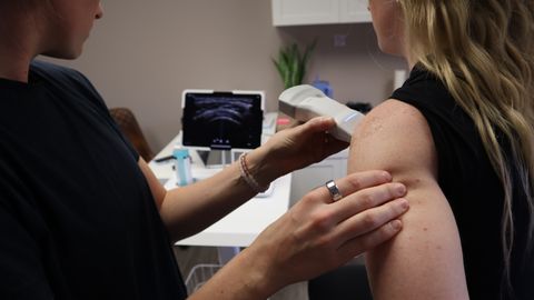
Ultrasound offers a dynamic view and can assess a tendon while it moves, which is useful for certain questions. Magnetic resonance imaging provides better contrast for surrounding structures and for tissue that ultrasound cannot reach easily. Neither is inherently superior, and the appropriate choice depends on the specific clinical question being asked.
The operator matters considerably as well, since ultrasound in particular depends on the skill of the person holding the probe. Comparing images taken on different machines or by different operators introduces variation that can easily be mistaken for genuine change.
Language shapes what athletes believe
Radiology reports use terminology developed for precision among clinicians, and that terminology lands very differently on an athlete. Words describing degeneration or tearing can suggest permanence and fragility that the clinical picture does not support.
How a finding is explained influences an athlete's willingness to load the region, which in turn influences the outcome. This is a recognised concern in sports medicine and is one reason results are increasingly discussed rather than simply handed over.
When imaging genuinely changes the plan
Imaging is most valuable when a specific diagnosis would alter management, such as distinguishing between conditions that need different approaches. It also matters where a serious alternative explanation needs to be excluded before an athlete resumes loading.
Outside those situations, an image often confirms what a competent examination has already established. The decision about whether to scan, and how to interpret what returns, belongs with the treating clinician who knows the athlete.
- Abnormal images are common in people without pain
- Structure and symptoms often move independently
- Imaging supports examination rather than replacing it